Biocracked
←

#ampk

2 articles
Approach: Macro science. Palette: B, warm.

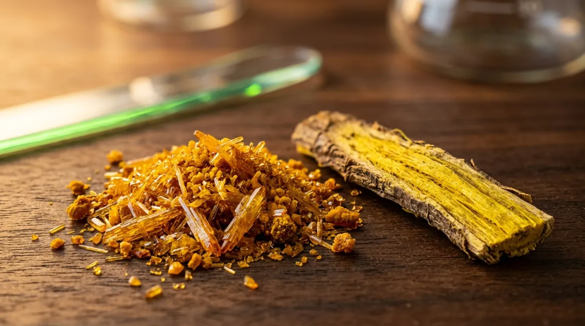
Extreme macro photograph of raw berberine: a small heap of vivid yellow-orange crystalline powder with needle-like crystal clusters, shot at a low 30-degree angle with a shallow depth of field. Beside it, a dry sliver of Berberis root bark shows its bright yellow inner wood and fibrous grain. The surface is a smooth dark walnut board. Low golden-hour side light from the left makes the crystals glint like amber glass and throws long warm copper shadows across the wood. One tiny electric green highlight reflects off a glass spatula edge in the soft background blur. The mood is quiet and investigative, with rich saturated honey and burnt-orange tones and no blue cast. Photorealistic, microscopy-like texture, no text or labels.
metabolism

Berberine vs. Metformin: The Pilot Trial Behind HbA1c 9.5% to 7.5%

BY HELIX WARD ·

A small 2008 pilot trial put berberine head-to-head with metformin in newly diagnosed type 2 diabetes, and the glucose-lowering effect looked similar. In the berberine group, HbA1c fell from 9.5% to 7.5% in three months. A second trial in 48 people already on treatment saw HbA1c drop from 8.1% to 7.3% and HOMA-IR fall 44.7%. But these were small studies in people with diabetes, and about a third of participants had GI side effects. Here is what the data show, what they don't, and what to know before you think about berberine.
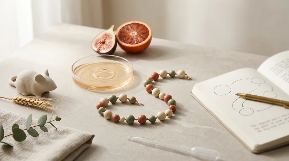
A wide, editorial-style still life photographed from slightly above on a pale warm-gray stone surface, lit by soft diffused morning light from the left. At the center, a delicate chain of 16 small, glossy beads in alternating muted terracotta, sage green, and cream is arranged in a gentle S-curve, each bead slightly different in size, like a hand-strung necklace. It stands for a peptide chain in a tactile, abstract way. Beside the chain, a translucent glass petri dish holds a thin layer of pale amber liquid with a few soft ripples. Behind it, a cross-section of a ripe fig and a halved blood orange show their intricate internal textures, suggesting cellular structure and mitochondria without literal illustration.

On the left side of the frame, a small ceramic mouse figurine in matte off-white sits calmly next to a sprig of dried wheat. It is a subtle nod to the mouse study and is stylized and minimal, not cartoonish. On the right, an open, softly blurred notebook with thin hand-drawn circles and curved lines, like looping pathway diagrams, lies under a brass pen. A few pages are visible but the text is illegible.

Scatter a few elements for texture: a small sprig of eucalyptus, a folded linen napkin in oatmeal tone, and a single clear glass pipette resting diagonally across the lower foreground. The palette is warm neutrals with accents of sage, terracotta, and soft amber, giving a calm, modern wellness-journal look. Shallow depth of field keeps the bead chain crisp while the background gently dissolves into creamy bokeh. Generous negative space across the upper right leaves room for headline overlay. The overall mood is clean, contemporary, and intellectually curious: scientific but warm, like a premium health and science magazine cover, with fine grain, natural shadows, and no text, logos, or futuristic elements.
peptides

MOTS-c Explained: The 16-Amino-Acid Mitochondrial Peptide That Activated AMPK and Shielded Mice From Diet-Induced Obesity

BY DR. SYNAPSE ·

A tiny 16-amino-acid peptide encoded in mitochondrial DNA, not the nucleus, activated AMPK in muscle and protected mice from diet-induced obesity and insulin resistance in a 2015 Cell Metabolism study. MOTS-c is now one of the most talked-about names in peptide and biohacking circles. But this was a mouse and molecular-discovery paper with no human outcome data, and MOTS-c is not an approved therapy. This article explains what MOTS-c is, how it appears to work through the folate cycle and AMPK, what the mice showed, and what readers should and should not conclude.

This page is available in