Biocracked
←

#mitoq

1 article
**Approach: Macro science. Palette: D, bold color field.**

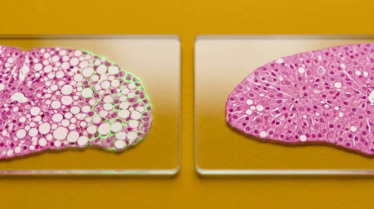
Extreme close-up microscopy-style photograph of two cross-sections of fatty mouse liver tissue, shown side by side on glass slides. Each slice is stained, with translucent pink-magenta tissue and pale, round fat droplets scattered through it. The left slice is dense with droplets and shows a faint electric green fluorescent glow along its cell edges. The right slice has clearer, more intact cell architecture and fewer droplets. The camera looks straight down at a slight tilt with a shallow depth of field, so the slide edges blur softly. Both slides lie on a saturated mustard-yellow backdrop. Crisp, even studio light casts soft, short shadows and gives the glass a clean reflection. The mood is analytical, comparative and precise, with a clear sense of two different outcomes.
peptides

MitoQ проти SS-31 у старих мишей з жирною печінкою: що вони зробили?

BY HELIX WARD ·

Два провідних препарати, націлених на мітохондрії — MitoQ та SS‑31 (elamipretide) — порівнювали в одній і тій же модельній дослідженні на мишах з жирною печінкою, і вони не замінювали один одного. Обидва знизили вміст жиру в печінці, зменшили запалення, пов’язане з NF‑κB та NLRP3, і покращили маркери інсулінового сигнального шляху. MitoQ виявився сильнішим антиоксидантом, тоді як SS‑31 краще зберігав структуру мітохондрій. Однак це дослідження проведено на старих самках мишей з різними дозуваннями та шляхами введення препаратів. Нижче — що показали результати, чого вони не доводять і чому слід бути обережним з ринком «дослідних» пептидів.

This page is available in