Biocracked
←

#mitoq

1 article
**Approach: Macro science. Palette: D, bold color field.**

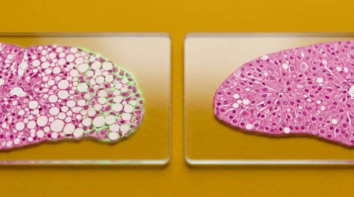
Extreme close-up microscopy-style photograph of two cross-sections of fatty mouse liver tissue, shown side by side on glass slides. Each slice is stained, with translucent pink-magenta tissue and pale, round fat droplets scattered through it. The left slice is dense with droplets and shows a faint electric green fluorescent glow along its cell edges. The right slice has clearer, more intact cell architecture and fewer droplets. The camera looks straight down at a slight tilt with a shallow depth of field, so the slide edges blur softly. Both slides lie on a saturated mustard-yellow backdrop. Crisp, even studio light casts soft, short shadows and gives the glass a clean reflection. The mood is analytical, comparative and precise, with a clear sense of two different outcomes.
peptides

노령 지방간 마우스에서 MitoQ와 SS-31, 각각 어떤 효과였을까?

BY HELIX WARD ·

두 주요 미토콘드리아 표적 화합물인 MitoQ와 SS‑31(엘라미프레타이드)을 같은 지방간 마우스 모델에서 비교한 결과, 두 물질은 서로 교환 가능한 것이 아니었다. 두 약물 모두 간 지방을 감소시키고 NF‑κB·NLRP3 염증을 억제했으며 인슐린 신호 전달 마커를 개선했다. MitoQ는 항산화 효과가 더 강했으며, SS‑31은 미토콘드리아 구조 보존에 더 뛰어났다. 다만 연구는 노령 암컷 마우스에 한정됐으며, 투여 용량·경로가 달라 직접적인 비교는 어렵다. 연구용 펩타이드 시장에 대한 경고도 함께 제시된다.

This page is available in