Biocracked
←

#mitoq

1 article
**Approach: Macro science. Palette: D, bold color field.**

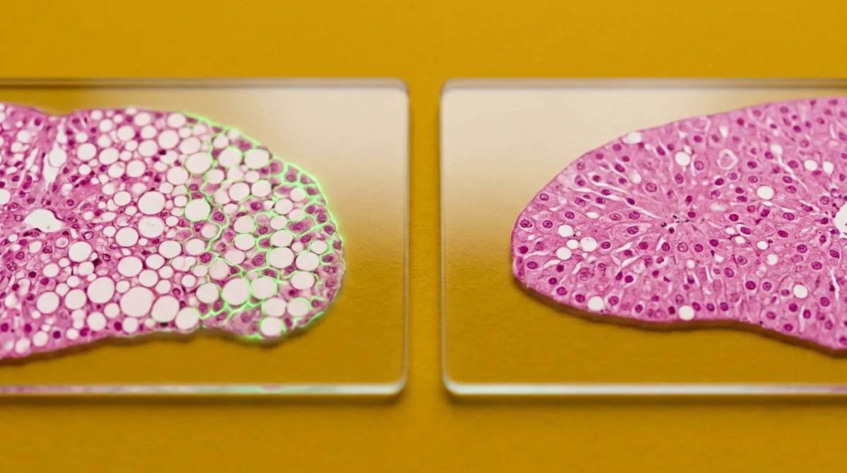
Extreme close-up microscopy-style photograph of two cross-sections of fatty mouse liver tissue, shown side by side on glass slides. Each slice is stained, with translucent pink-magenta tissue and pale, round fat droplets scattered through it. The left slice is dense with droplets and shows a faint electric green fluorescent glow along its cell edges. The right slice has clearer, more intact cell architecture and fewer droplets. The camera looks straight down at a slight tilt with a shallow depth of field, so the slide edges blur softly. Both slides lie on a saturated mustard-yellow backdrop. Crisp, even studio light casts soft, short shadows and gives the glass a clean reflection. The mood is analytical, comparative and precise, with a clear sense of two different outcomes.
peptides

MitoQ vs SS-31 in Aged Fatty-Liver Mice: Which Did What?

BY HELIX WARD ·

Two leading mitochondria-targeted compounds, MitoQ and SS-31 (elamipretide), were compared in the same mouse model of fatty liver, and they were not interchangeable. Both cut liver fat, dampened NF-κB and NLRP3 inflammation, and improved insulin-signaling markers. MitoQ showed the stronger antioxidant signature, while SS-31 did better at preserving mitochondrial structure. But this was a study in aged female mice, with different doses and routes for each drug. Here is what the results show, what they don't, and why the research-peptide market deserves caution.

This page is available in