Biocracked
←

#subcutaneous-fat

2 articles
**Approach 1 (macro science), Palette D (bold color field).**

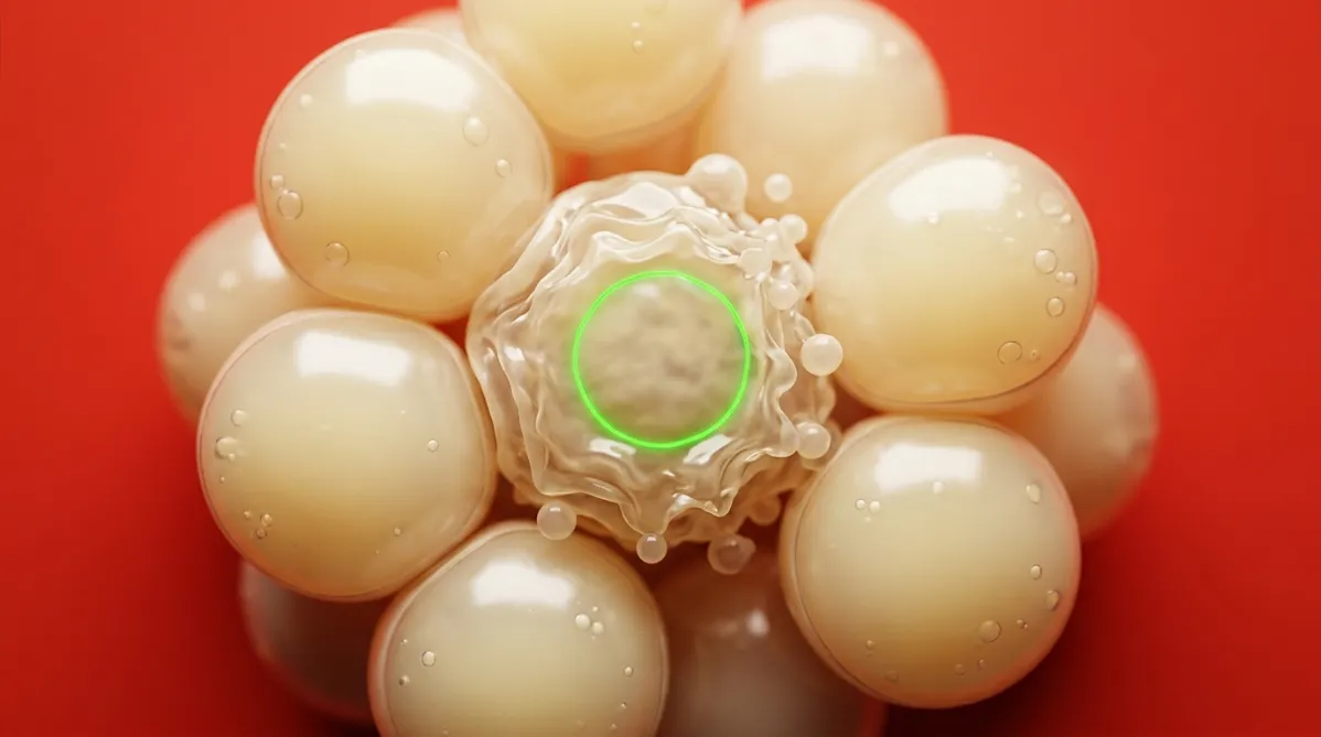
Extreme close-up microscopy-style photograph of a cluster of fat cells (adipocytes), rendered as translucent, pearly spheres with thin membranes and tiny lipid droplets inside. One cell in the center is visibly shrinking and blebbing, its membrane crinkled and fragmenting into small beads, the look of apoptosis. A few of the surrounding cells are intact and glossy. The cells sit against a saturated tomato-red backdrop, so the pale yellow-cream spheres stand out sharply. The camera looks straight down at shallow depth of field, with the focal plane on the dying cell and the edges softly blurred. Soft, diffuse top light gives gentle highlights and warm shadows, with no blue cast. One thin electric green fluorescent ring around the central cell's nucleus is the only green accent. The mood is analytical, precise and quietly skeptical.
research

Hoe werkt CBL-514? De wetenschap uitgelegd

BY DR. SYNAPSE ·

CBL-514 wordt niet gepresenteerd als een vet‑smelt‑ of vet‑verbrandingsmiddel. De bewering is dat het de overlevingskinase DYRK1b blokkeert en vetcellen richting apoptose duwt via caspase 3 en Bax/Bcl‑2. Dit bewijs komt uit niet‑gepubliceerde bedrijfsrapporten die in de fase‑2a‑ en fase‑2‑publicaties worden genoemd, en de chemische structuur is niet bekendgemaakt. De fase‑2‑studie uit 2025 meette vetvolume met echografie, foto’s en veiligheid, zonder menselijke biopsies of caspase‑data. Dit artikel scheidt de moleculaire hypothese van wat daadwerkelijk bij mensen is gemeten, inclusief de hoge uitval en single‑blind opzet.
Approach 1, macro science, in palette D, bold color field.

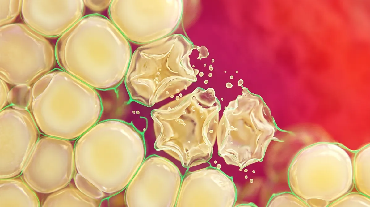
Extreme close-up photograph of a cluster of fat cells (adipocytes) in tissue, shot like a stained microscopy image. Rounded, translucent, pearly-yellow lipid-filled cells press together in a honeycomb. Several in the center look collapsed and shriveled, with ruptured membranes and scattered droplets. A few fine threads of electric green (#00e676) fluorescent stain outline the cell walls. The camera is straight-on at shallow depth of field, with the background cells melting into soft blur. Bright, even transmitted light makes the membranes glow. The backdrop is a saturated magenta-to-tomato-red field that sets off the pale yellow cells. The mood is scrutinizing, skeptical and precise. No text, no labels.
research

CBL-514 vetverwijderende injecties: wat zegt de wetenschap?

BY DR. SYNAPSE ·

CBL‑514 heeft nu een placebogecontroleerde fase‑2‑gerandomiseerde proef in *Aesthetic Surgery Journal*, waarin de auteurs melden dat zowel de primaire als de secundaire eindpunten zijn behaald. Deze beoordeling laat echter zien waarom het onderzoek kritisch bekeken moet worden: de studie was enkel‑blind, terwijl onderzoekers en sponsor niet geblindeerd waren; bovendien heeft de sponsor meegewerkt aan ontwerp, analyse en manuscript. Van de 75 behandelde deelnemers voltooiden er slechts 38 de studie en de per‑protocol‑groep bestond uit slechts 30 personen. De follow‑up eindigde acht weken na de laatste behandeling en de deelnemers waren overwegend vrouwen en witte personen. Hieronder lees je wat de proef wel en niet kan aantonen, en waarom dit geen medisch advies of bewijs van een goedgekeurde behandeling is.

This page is available in