Biocracked
←

#subcutaneous-fat

2 articles
**Approach 1 (macro science), Palette D (bold color field).**

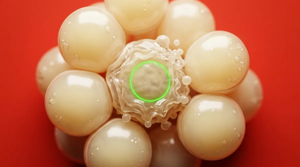
Extreme close-up microscopy-style photograph of a cluster of fat cells (adipocytes), rendered as translucent, pearly spheres with thin membranes and tiny lipid droplets inside. One cell in the center is visibly shrinking and blebbing, its membrane crinkled and fragmenting into small beads, the look of apoptosis. A few of the surrounding cells are intact and glossy. The cells sit against a saturated tomato-red backdrop, so the pale yellow-cream spheres stand out sharply. The camera looks straight down at shallow depth of field, with the focal plane on the dying cell and the edges softly blurred. Soft, diffuse top light gives gentle highlights and warm shadows, with no blue cast. One thin electric green fluorescent ring around the central cell's nucleus is the only green accent. The mood is analytical, precise and quietly skeptical.
research

Bagaimana cara kerja CBL‑514? Inilah ilmunya

BY DR. SYNAPSE ·

CBL‑514 tidak diposisikan sebagai obat pencair lemak atau pembakar lemak. Klaim yang diajukan adalah bahwa ia menghambat kinase kelangsungan hidup DYRK1b dan mendorong sel lemak menuju apoptosis melalui caspase‑3 serta rasio Bax/Bcl‑2. Bukti tersebut berasal dari laporan perusahaan yang belum dipublikasikan dan dikutip dalam makalah fase 2a serta fase 2, sementara struktur kimianya belum diungkapkan. Uji fase 2 tahun 2025 mengukur volume lemak dengan ultrasonografi, foto, dan data keamanan, tanpa biopsi manusia ataupun data caspase. Artikel ini memisahkan hipotesis molekuler dari apa yang sebenarnya diukur pada manusia, termasuk tingkat drop‑out yang tinggi dan desain single‑blind.
Approach 1, macro science, in palette D, bold color field.

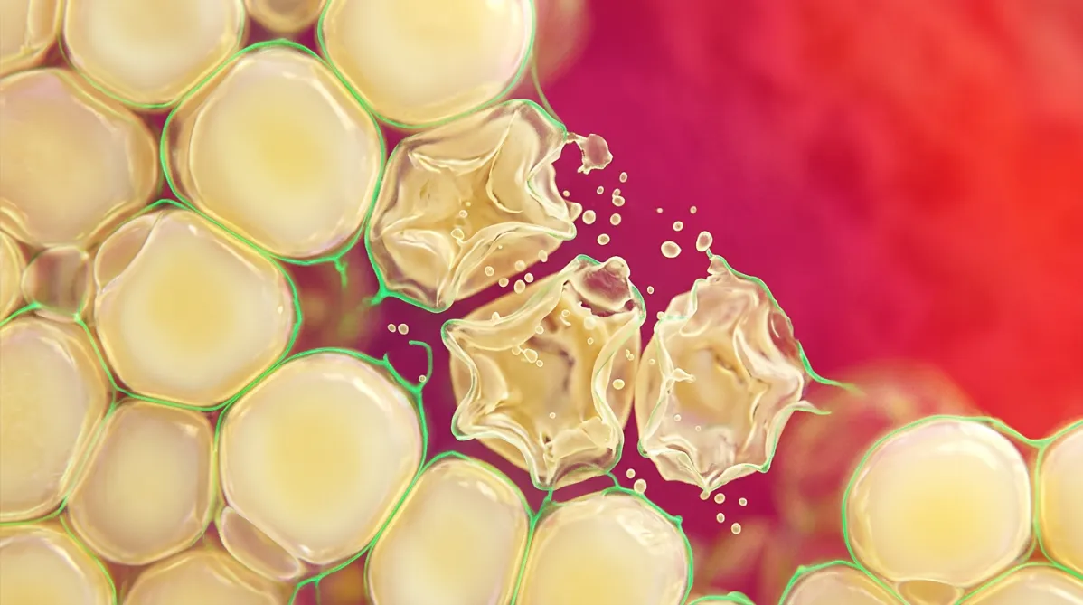
Extreme close-up photograph of a cluster of fat cells (adipocytes) in tissue, shot like a stained microscopy image. Rounded, translucent, pearly-yellow lipid-filled cells press together in a honeycomb. Several in the center look collapsed and shriveled, with ruptured membranes and scattered droplets. A few fine threads of electric green (#00e676) fluorescent stain outline the cell walls. The camera is straight-on at shallow depth of field, with the background cells melting into soft blur. Bright, even transmitted light makes the membranes glow. The backdrop is a saturated magenta-to-tomato-red field that sets off the pale yellow cells. The mood is scrutinizing, skeptical and precise. No text, no labels.
research

Injeksi Penghapusan Lemak CBL-514: Apa Kata Ilmu?

BY DR. SYNAPSE ·

CBL-514 kini memiliki uji coba acak fase 2 yang dikontrol plasebo dalam *Aesthetic Surgery Journal*. Penulis melaporkan bahwa uji coba tersebut memenuhi endpoint primer dan sekunder, namun desainnya perlu diteliti secara kritis: uji coba bersifat single‑blind dengan peneliti dan sponsor yang tidak diblind, serta sponsor terlibat dalam perancangan, analisis, dan penulisan studi. Dari 75 peserta yang menerima perlakuan, hanya 38 yang menyelesaikan, dan kelompok per‑protokol hanya berisi 30 orang. Tindak lanjut berakhir 8 minggu setelah perlakuan terakhir, dengan sampel mayoritas perempuan dan berkulit putih. Berikut apa yang dapat didukung oleh uji coba ini, apa yang tidak, serta mengapa ini bukan saran medis atau bukti perawatan yang disetujui.

This page is available in